Ultrasound technology is a medical imaging technique that uses high-frequency sound waves to create images of the inside of the body.
It is commonly used to visualize muscles, tendons, blood vessels, and organs such as the heart, liver, kidneys, and reproductive organs.
How Ultrasound Works:
Sound Waves:
Ultrasound uses sound waves that are beyond the frequency range of human hearing (typically 20,000 Hz or higher).
These sound waves are emitted by a device called a transducer, which is placed on the skin or inserted into the body in some cases.
Echoes:
As the sound waves travel through the body, they bounce (or echo) off structures inside the body.
These echoes are then captured by the transducer.
Image Creation:
The ultrasound machine processes the sound wave echoes and converts them into real-time images or videos, which can then be interpreted by medical professionals.
Applications of Ultrasound
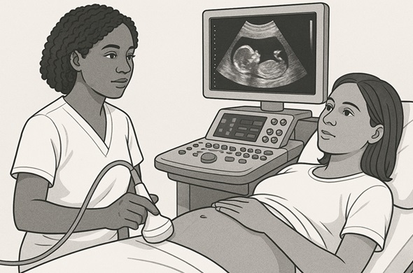
Obstetrics and Gynaecology
Pregnancy: Ultrasound is commonly used to monitor fetal development, check for abnormalities, and determine the due date.
Pelvic Exam:Used to assess organs like the uterus and ovaries.
Cardiology
Echocardiography: Ultrasound is used to create images of the heart, enabling doctors to evaluate its size, shape, and function.
Abdominal Imaging
Commonly used to examine the liver, gallbladder, kidneys, spleen, and pancreas for abnormalities, such as tumours or stones.
Musculoskeletal Imaging
Ultrasound can assess joints, muscles, tendons, and ligaments for tears, inflammation, or injuries.
Vascular Ultrasound
Used to assess blood flow in veins and arteries. It helps diagnose conditions such as deep vein thrombosis (DVT) or arterial blockages.
Guided Procedures
Ultrasound is often used to guide needle placements for biopsies, injections, or catheter insertions, ensuring accuracy.
Advantages of Ultrasound
Non-invasive:No need for incisions or injections in most cases.
Real-time Imaging:Provides live images of the body’s internal structures.
Safe:Unlike X-rays or CT scans, ultrasound does not involve ionizing radiation, making it a safer option, especially for pregnant women.
Portable and Cost-effective: Compared to other imaging technologies, ultrasound equipment is relatively inexpensive and can be used in a variety of settings, including at the point of care.
Limitations of Ultrasound
Image Quality:While ultrasound provides detailed images, it may not be as clear or precise as other imaging techniques like CT scans or MRIs.
Operator Dependence: The quality of the images depends on the skill of the technician performing the ultrasound.
Limited Penetration: Ultrasound is less effective for imaging certain areas of the body, such as the lungs or structures behind bone.
Recent Advancements
3D and 4D Ultrasound:These advanced ultrasound technologies provide three-dimensional images, which are especially popular in obstetrics to give a more detailed view of the fetus.
Elastography:This technique assesses tissue stiffness, which is helpful for detecting liver fibrosis or cancer.
Portable Ultrasound Devices:Miniaturized ultrasound machines have been developed, allowing more widespread use in remote areas or emergency settings.


 FAQ
FAQ Contact Us
  New Batch : 9555124124/ 7428085757
  Tech Support : 9555124124/ 7428085757